I used to think “3D bioprinting” was just a cool sci-fi phrase that tech people liked to throw around at conferences. Then I watched a researcher print living tissue on a lab bench, and it felt less like science fiction and more like a very rough, very real prototype of the future.
If you only want the short version: we are not printing full, ready-to-transplant human hearts or kidneys yet. We are, however, printing simpler tissues, small organ-like structures, and surgical grafts that are already helping patients in limited ways. Over the next 10 to 20 years, 3D bioprinting will likely move from lab experiments to real transplant options for certain organs, but the big, complex organs will probably come last and require heavy regulation and long-term testing.
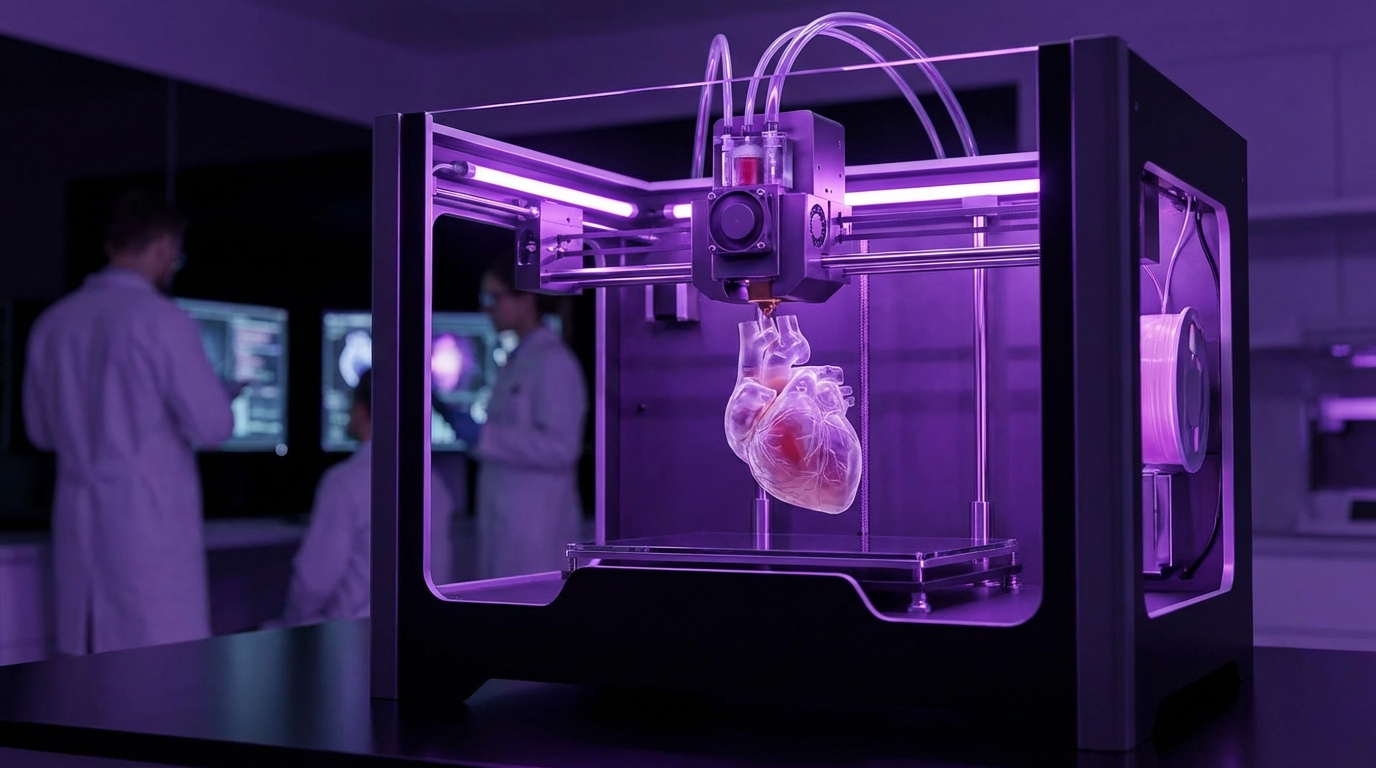
What 3D bioprinting actually is (beyond the buzzword)
If you have used a regular 3D printer before, the basic idea is familiar. A machine moves in 3 dimensions and deposits material layer by layer to build an object.
With 3D bioprinting, that object is living tissue, and the material is “bioink” instead of plastic.
3D bioprinting is the process of layer-by-layer printing of living cells and biomaterials to create tissue-like structures that can grow, integrate, or function inside the body.
The catch: living cells behave nothing like melted plastic. They die if conditions are wrong. They migrate. They divide. They communicate. That brings a whole set of technical and ethical puzzles.
How 3D bioprinting works at a high level
The workflow usually looks like this:
- Start with a digital model of the tissue or organ (from MRI/CT scans or computer design).
- Prepare bioinks: mixtures of living cells and supportive materials (hydrogels, proteins, polymers).
- Print the structure layer by layer with a bioprinter.
- Culture the printed construct in a bioreactor so the cells mature and organize.
- Test for function, safety, and stability before any thought of transplant.
It sounds clean and linear. In practice, every step fights back.
Main bioprinting technologies you keep hearing about
Once you start reading papers, three broad printing methods show up again and again:
- Extrusion bioprinting
Think of a syringe squeezing out toothpaste. A mechanical or pneumatic system pushes cell-laden bioink through a nozzle. It is slow and relatively gentle, good for thick tissues, but the resolution is limited. - Inkjet bioprinting
Similar to an inkjet printer head. Tiny droplets of cell suspensions are ejected in controlled patterns. It is faster and can have higher resolution, but the droplets must be low viscosity, and cells can get damaged by heat or pressure. - Light-based bioprinting (stereolithography, digital light processing)
A light source cures photosensitive bioinks in a pattern. You get very fine structures and smoother surfaces, but you need bioinks that respond well to light without harming the cells.
Each method forces tradeoffs between precision, speed, cell survival, and what kinds of tissues you can build.
What is inside a “bioink” anyway?
“Bioink” sounds fancy, but you can break it down into two parts:
| Component | Role | Examples |
|---|---|---|
| Cells | Provide the living, functional part of the tissue | Stem cells, patient-derived cells, organ-specific cells (hepatocytes, cardiomyocytes, etc.) |
| Matrix / Hydrogel | Provide structure and a local environment for the cells | Collagen, gelatin, alginate, fibrin, synthetic polymers like PEG-based gels |
The hard part is that these inks must be:
- Printable (they should hold shape during and after deposition).
- Biocompatible (they should keep cells alive and happy).
- Degradable or remodelable (the body needs to replace them with its own matrix over time).
- Strong enough for surgery (at least for certain organs or grafts).
You push one property up, and another tends to drop. You want more strength? You risk harming cell viability. You want more cell-friendly softness? The printed construct might collapse.
The central tension in bioprinting is balancing “good engineering plastic” behavior with “good living tissue” behavior in the same material.
Where 3D bioprinting is helping patients right now
So, are hospitals printing hearts in the basement? No.
What you do see are more modest, but still meaningful, uses.
Custom surgical models and guides
Technically, these are often printed with regular 3D printers, not bioprinters, but they ride on the same digital pipeline.
The workflow:
- Scan the patient (CT or MRI).
- Convert images to a 3D model.
- Print a physical model of the organ or bone with plastic.
- Use it for surgical planning or as a guide in the operating room.
This is not living tissue, but it is part of the same bigger story: turning medical imaging into physical, patient-specific objects.
Bioprinted skin and cartilage
Skin and cartilage are much more forgiving than, say, a liver.
They have:
- Simple architecture (fewer cell types than complex organs).
- No need for immediate deep vascular networks in the same way as larger organs.
- Lower metabolic demand.
Real-world progress:
- Skin grafts for burns: Companies and research groups print layered skin constructs (epidermis + dermis) and test them for wound coverage. Some early clinical applications focus on improving healing and reducing scarring.
- Cartilage for ears and noses: Bioprinted cartilage is being explored to repair defects or reconstruct parts of the ear or nose with a custom shape based on the patient.
These are not yet mass-market products, but they are well ahead of full organ bioprinting.
Tracheal and vascular grafts
There have been high-profile cases where surgeons implanted printed tracheal splints or grafts that support a child’s airway. Some of these devices are made from synthetic materials; others are mixed with cells or coated to encourage tissue integration.
Blood vessels are another focus:
A fully printed organ is useless without a working vascular network to feed it.
Researchers are printing:
- Small vessel networks inside hydrogels.
- Vascular channels that can later be seeded with endothelial cells.
- Scaffolds that guide natural vessel growth into a printed tissue.
These first steps are less about transplant tomorrow and more about solving basic infrastructure problems that all big organs share.
Bioprinted organs vs “organoids” vs organ-on-a-chip
This part gets confusing because the terms sound similar.
Organoids and mini-organs
Organoids are small, self-organizing cell clusters grown from stem cells. They mimic some features of real organs, such as:
- Liver organoids with detox and protein-synthesis functions.
- Brain organoids forming primitive neural networks.
- Intestinal organoids with villus-like structures.
Researchers sometimes use bioprinting to place organoids inside a structured scaffold or to combine them in specific patterns. The result is a kind of “enhanced” organoid.
These mini-organs are not ready for transplant, but they already matter for:
- Drug testing and toxicity studies.
- Studying genetic diseases.
- Exploring how cells interact in a 3D microenvironment.
Organ-on-a-chip systems
Organ-on-a-chip devices use microfluidic channels and tiny tissue constructs to model parts of organ function on a small scale.
Bioprinting helps by:
- Placing cells with high precision inside the chip.
- Creating layered barriers (like lung air-blood interfaces).
- Integrating vascular-like channels.
Here again, the goal is not transplant. It is prediction: how will a drug behave in human tissue before you give it to an actual human.
Where true transplantable organs fit in
You can think of it this way:
| Category | Scale | Main Use Today | Transplant Potential |
|---|---|---|---|
| Organoids | Millimeter to a few centimeters | Research, disease modeling, drug testing | Long-term, maybe as building blocks |
| Organ-on-a-chip | Micro to millimeter | Drug testing, toxicity screening | No direct transplant, but informs design |
| Bioprinted tissues | Centimeter to organ scale | Early grafts, experimental therapy | Target for transplant work |
So when someone asks, “Can we print a kidney and just plug it in?”, the honest reply is: we are still learning how to print a small, stable, well-perfused piece of kidney-like tissue that functions reliably in an animal.
Why printing a full organ is so hard
At a high level, you face at least five big hurdles. There are more, but these five repeat across every organ.
1. Vascularization: keeping tissue alive beyond a few millimeters
Living cells need oxygen and nutrients. Diffusion alone only works across short distances. Past a certain thickness, you need blood vessels or the inner cells die.
Bioprinting tries to solve this through:
- Direct printing of vessel-like channels: Printing empty channels that you later seed with endothelial cells.
- Sacrificial inks: Printing a sacrificial material inside a hydrogel, then dissolving it to leave hollow channels.
- Angiogenic factors: Embedding growth factors that encourage the host body to grow vessels into the graft.
The problem is not just making channels. They must:
- Connect properly to the host’s vessel tree.
- Stay open and not collapse.
- Resist clotting.
- Grow and remodel over time.
In a way, printing the vessels is harder than printing the bulk of the organ itself.
Without a stable vascular network, a printed organ is a beautiful sculpture that dies from the inside out.
2. Tissue organization and microarchitecture
Organs are not just blobs of cells.
A liver has lobules with specific orientation. A kidney has glomeruli, tubules, and a precise filtration layout. A heart has aligned muscle fibers and conduction pathways.
Bioprinting can place different cell types in patterns, but true self-organization still relies on biology.
Questions researchers are still wrestling with:
- How much structure should we print, and how much should we let the cells organize themselves?
- How do we print gradients (of stiffness, chemical cues, oxygen) that guide this self-organization?
- Can we recreate development-like pathways in a mature body safely?
This is where the line between “engineering project” and “developmental biology experiment” gets blurry.
3. Mechanical strength and durability
Organs face physical stress:
- Hearts contract billions of times.
- Lungs expand and contract with every breath.
- Joints carry body weight.
Hydrogels and soft matrices are kind to cells but often mechanically weak. Stronger synthetic materials may harm cells or trigger immune responses.
Finding materials that:
- Survive surgical handling.
- Maintain shape and function under load.
- Degrade at a controlled rate.
is not trivial.
Some teams are combining:
- Printed cell-laden soft regions for function.
- Printed or prefabricated stiffer frameworks for support.
You end up with hybrid constructs that look less like a printed block and more like a composite device.
4. Immune compatibility and rejection
3D bioprinting is often presented as an automatic fix for organ rejection: “We will just print organs from the patient’s own cells, so no immune problems.”
Reality is more complicated.
Issues:
- Patient-derived cells are not always available or healthy, especially in older or very sick people.
- The biomaterials used in the matrix may still trigger an immune response.
- Cancer risk must be controlled if stem cells are used.
- If gene editing is applied to cells, that adds another layer of safety and ethics review.
There is a spectrum of approaches:
| Cell Source | Pros | Cons |
|---|---|---|
| Autologous (patient’s own) | Lower rejection risk, better compatibility | Time-consuming, costly, may be poor quality cells |
| Allogeneic (donor or universal lines) | Off-the-shelf, faster, more standardized | Higher rejection risk, imagine lifelong immunosuppression |
| Gene-edited “universal” cells | Potentially broad use with fewer immune reactions | Regulatory and ethical complexity, long-term safety unknown |
3D bioprinting does not sidestep immunology; it pulls immunology right into the center of organ manufacturing.
5. Regulatory, quality, and manufacturing scale
Let us say, hypothetically, a team figures out how to print a functional piece of liver tissue and shows it works in animals.
You still need to answer:
- How do you prove safety and consistent quality to regulators like the FDA or EMA?
- How do you manufacture these constructs under strict standards (GMP) at scale?
- How do you track every cell lot, every printing run, every material batch?
Unlike a pill, a printed organ is:
- Complex.
- Biologically active.
- Personalized (in many scenarios).
That makes standardization hard. Every deviation in the process could change the risk profile.
Printing one working organ in a lab is a scientific achievement. Printing thousands safely and consistently is a manufacturing and regulatory challenge.
Which organs will likely arrive first?
I do not think all organs are equal candidates. Some will hit clinical use earlier because they are structurally simpler or can work as partial grafts instead of full replacements.
Good near- to mid-term candidates
- Skin: Already moving from lab to limited clinical use. More advanced, vascularized skin constructs are in active development.
- Cartilage (ear, nose, joint surfaces): Structurally simpler, no heavy vascularization, already in preclinical and early clinical work.
- Bone segments: Bioprinted bone grafts with living cells and growth factors, especially for non-load-bearing or partially load-bearing scenarios.
- Small vascular grafts: Replacing sections of damaged vessels, especially where synthetic grafts fail.
- Cornea-like constructs: Transparent, layered tissues for certain eye repairs.
These do not solve the full organ shortage, but they address real clinical needs.
More distant but extremely valuable targets
- Liver: A partial liver construct that provides enough support while the native liver recovers, or as a bridge to transplant, is a serious active goal.
- Kidney: Chronic kidney disease and dialysis drive huge demand. Even a partial kidney-like device that handles certain aspects of filtration could be powerful.
- Pancreatic tissue: Insulin-producing cell constructs for diabetes are already in trials, often encapsulated rather than fully printed organs.
I would not expect a fully functional, fully printed human heart or kidney widely available in standard clinical practice in the next few years. A decade or two is more reasonable, with many caveats.
Ethical questions that will not go away
The technology side often gets all the attention, but the ethics are just as tricky.
Who gets access, and at what price?
If 3D bioprinted organs become real, they will be:
- Complex to produce.
- Heavily regulated.
- Likely expensive at first.
So you run into:
- Equity: Do only wealthy patients or rich countries benefit originally?
- Prioritization: Which conditions and which patients are treated first?
- Insurance and reimbursement: Who pays for a one-off custom organ?
It is very easy to say “this will save lives,” and it likely will. But without careful policy, it can also widen gaps in health outcomes.
Where do the cells come from?
If patient-specific cells are not always practical, cell banks and universal donors enter the picture.
Questions:
- How are donors recruited, consented, and compensated?
- How is genetic privacy handled if cell lines are stored long term?
- Can commercial companies own key cell lines that become widely used?
And if we start editing those cells for compatibility or function, we walk into gene-editing ethics as well.
Upgrading vs restoring
Right now, the goal is to restore lost or failing function. But in theory, printed tissues could be:
- Stronger.
- More durable.
- Resistant to certain diseases.
Then you run into questions like:
- Is a slightly “improved” organ acceptable if it saves a life?
- Where is the line between therapy and enhancement?
- Do we treat enhancements differently from replacements in regulation and insurance?
I do not think these questions have clear answers yet. But waiting until the technology is mature to talk about them is too late.
Where AI and software intersect with 3D bioprinting
Since your niche is technology, you might be curious where AI shows up in all this.
Designing organs computationally
Bioprinting starts from a digital model. For complex organs, these models are not just shapes. They encode:
- Vascular trees.
- Mechanical property maps.
- Cell-type distributions.
AI and simulation tools can help:
- Generate candidate designs for vascular networks that maintain perfusion.
- Predict mechanical stresses and guide material placement.
- Optimize print paths for cell viability and print speed.
“Generative design” in engineering has a medical cousin here. The computer may propose vessel layouts that a human would not think of, then you simulate and refine.
Monitoring prints and predicting failures
Real-time monitoring matters. During printing, you can track:
- Nozzle pressure and flow.
- Layer shape through cameras or sensors.
- Temperature, humidity, and other environmental variables.
Machine learning can then:
- Detect deviations from normal patterns in real time.
- Predict when a print is likely to fail.
- Recommend adjustments during the run.
You can think of it as quality control with built-in prediction.
Modeling tissue growth after printing
After printing, cells:
- Migrate.
- Divide.
- Remodel the matrix.
AI-driven models can simulate that behavior over time, helping teams:
- Predict how a construct will change structurally.
- Estimate when it is ready for implantation.
- Anticipate weak spots or regions prone to necrosis.
It is not perfect, but even rough predictions can save months of trial-and-error experiments.
Bioprinting is physical, but the design, monitoring, and prediction layers are increasingly software-intensive and data-heavy.
What this means if you are building in healthtech or medtech
If you work or plan to work in health-related tech, you do not have to print the organ yourself to be relevant to this field.
Practical opportunity areas around bioprinting
- Imaging and segmentation tools: Better pipelines that take CT/MRI data and generate clean, printable 3D models with minimal manual editing.
- Simulation software: Tools that help researchers or clinicians simulate blood flow, mechanical stresses, or diffusion in printed constructs.
- Lab automation: Systems that coordinate bioreactors, printers, and quality control steps with reliable data logging.
- Data management and traceability: Platforms for tracking cell batches, material lots, print logs, and patient data in a compliant way.
- Predictive analytics for process control: Models that learn from past print runs and guide parameter selection for new ones.
These areas are far less glamorous than “we printed a heart,” but they are necessary layers if bioprinting is going to scale.
Where I would be careful about hype
You will see bold claims like “organs on demand in 5 years” or “no more donor waiting lists soon.” I would not build a business plan on that timeline.
Questions to ask when you see a big claim:
- Was the result shown in vitro, in small animals, in large animals, or in humans?
- Was it a partial tissue or a full functional organ?
- How long did the construct survive and perform?
- Was this a one-off research demo or part of a controlled trial?
- Is there a clear regulatory strategy mentioned anywhere?
If the answer to most of these is vague, the claim is probably ahead of the data.
Where we realistically are, and what to expect next
To ground everything:
Right now, 3D bioprinting is reliably producing simple tissues and research models, and it is starting to touch clinical care with grafts and specialized constructs, but it is still far from routine printing of full human organs for transplant.
Here is a rough, non-rigid sense of progress across application types:
| Application | Stage | Examples |
|---|---|---|
| Surgical models (plastic) | Clinical routine in many centers | Cardiac models, jaw reconstruction planning |
| Bioprinted skin, cartilage | Preclinical and limited clinical trials | Burn treatments, ear/nose reconstruction |
| Organ-on-a-chip, organoids with printing | Research and some industry use | Drug safety testing, disease modeling |
| Partial organ grafts (liver, pancreas segments) | Preclinical animal studies | Liver support constructs, islet-like tissues |
| Full solid organ replacement | Concept and early animal research | Prototype hearts, kidneys in large animals |
If you care about the technology side, the next 5 to 10 years will likely be about:
- More standardization in printers and bioinks.
- Better vascularization strategies.
- Clearer regulatory paths for early therapies.
- Deeper integration of imaging, AI, and bioprinting workflows.
I will be surprised if every prediction about timelines turns out right. Some problems might fall faster than expected. Others may drag for decades. Biology is stubborn.
But the direction of travel is quite clear: from static, generic implants toward living, patient-specific, and eventually printed tissues that actually participate in the body’s own repair processes.